Applications:
CYTOCOLOR stain kits can help you make a diagnosis of hematologic disorders. Here are only a few of them ...
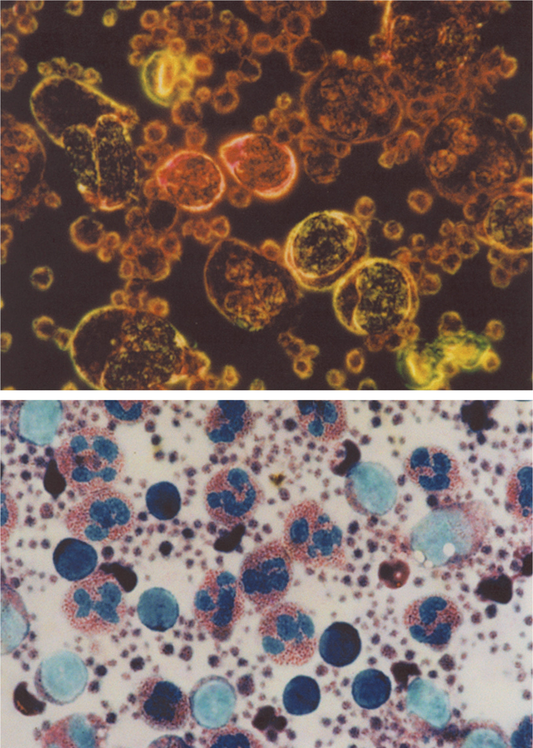
- Iron deficiency anemia: PANOPTIKON or RAPIDIFF visualize nuclear and cytoplasmic features of erythroblasts, including defective hemoglobinization.
- Autoimmune hemolytic anemia: PANOPTIKON or RAPIDIFF distinguish polychromatophilic erythrocytes from normal erythrocytes, and stains erythroblasts vividly.
- Megaloblastic anemia: NEUTROCOLOR demonstrates neutrophils that show distinctive crimson red granulation. GRANULOCOLOR reveals a pink color in neutrophil granules. PANOPTIKON or RAPIDIFF display nuclear and cytoplasmic features of megaloblasts and atypical granulocytes.
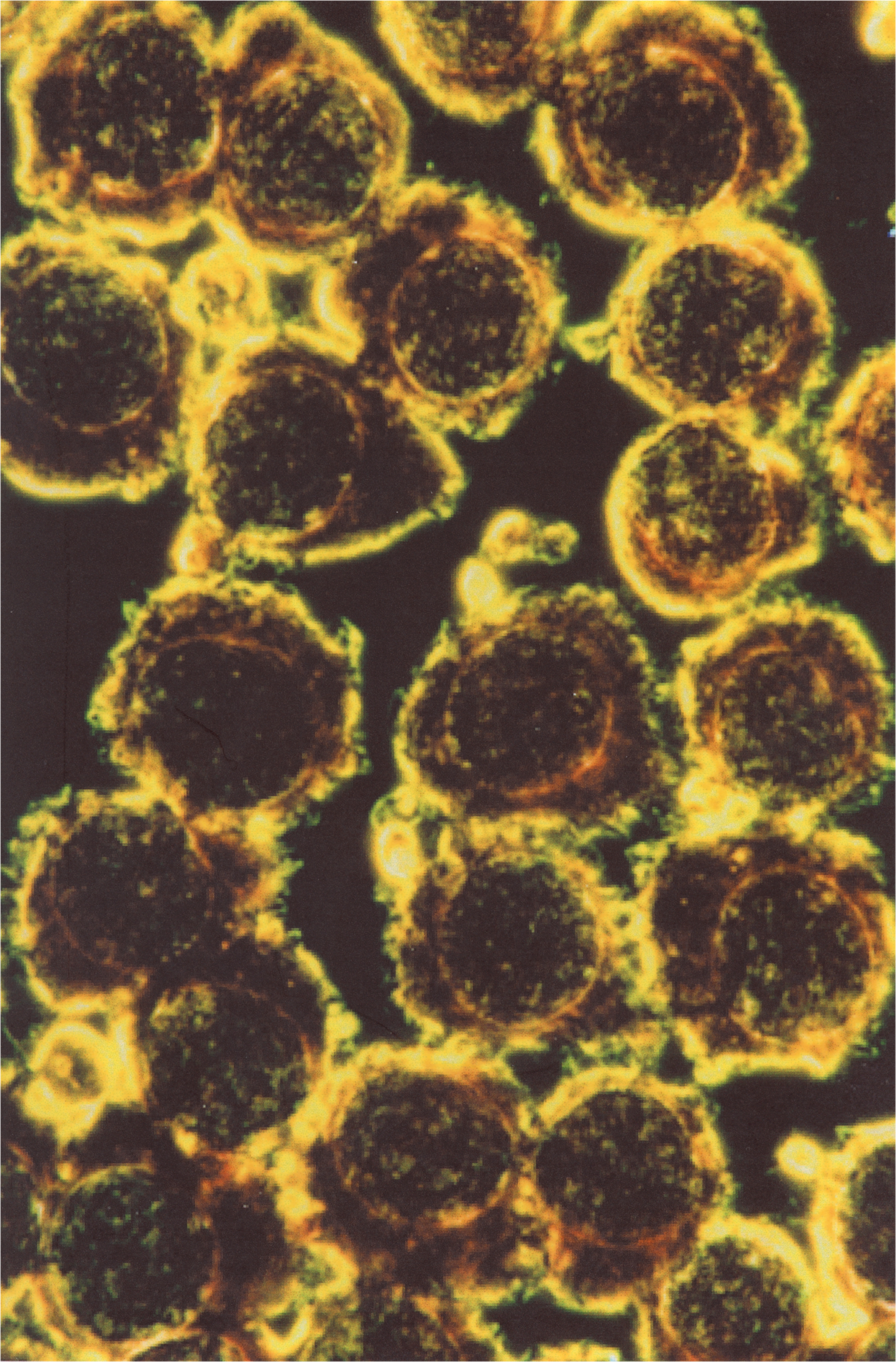
- Chronic granulocytic leukemia: RAPIDIFF or PANOPTIKON display vivid details of nuclear and cytoplasmic structures. In basophils, granules stain bright red. LYSOCOLOR demonstrates lysosomes in immature cells such as promyelocytes and myeloblasts. GRANULOCOLOR visualizes granules in all types of granulocytic cells. Using MEGACOLOR, dwarf megakaryocytes can be identified.
- Acute myeloblastic leukemia: Using LYSOCOLOR, granules in myeloblasts and promyelocytes stain black, as do Auer rods. With GRANULOCOLOR, both primary and secondary granules are visualized.
- Acute promyelocytic leukemia: Using LYSOCOLOR, granules in leukemic blasts are numerous and stain black. Auer rods also stain black. With GRANULOCOLOR, Auer rods stain red.
- Myelodysplastic syndrome: In refractory anemia with excess blasts (RAEB), lysosomes in myeloblasts stain black with LYSOCOLOR, and details of nuclear chromatin and cytoplasm are distinct after staining with PANOPTIKON or RAPIDIFF.
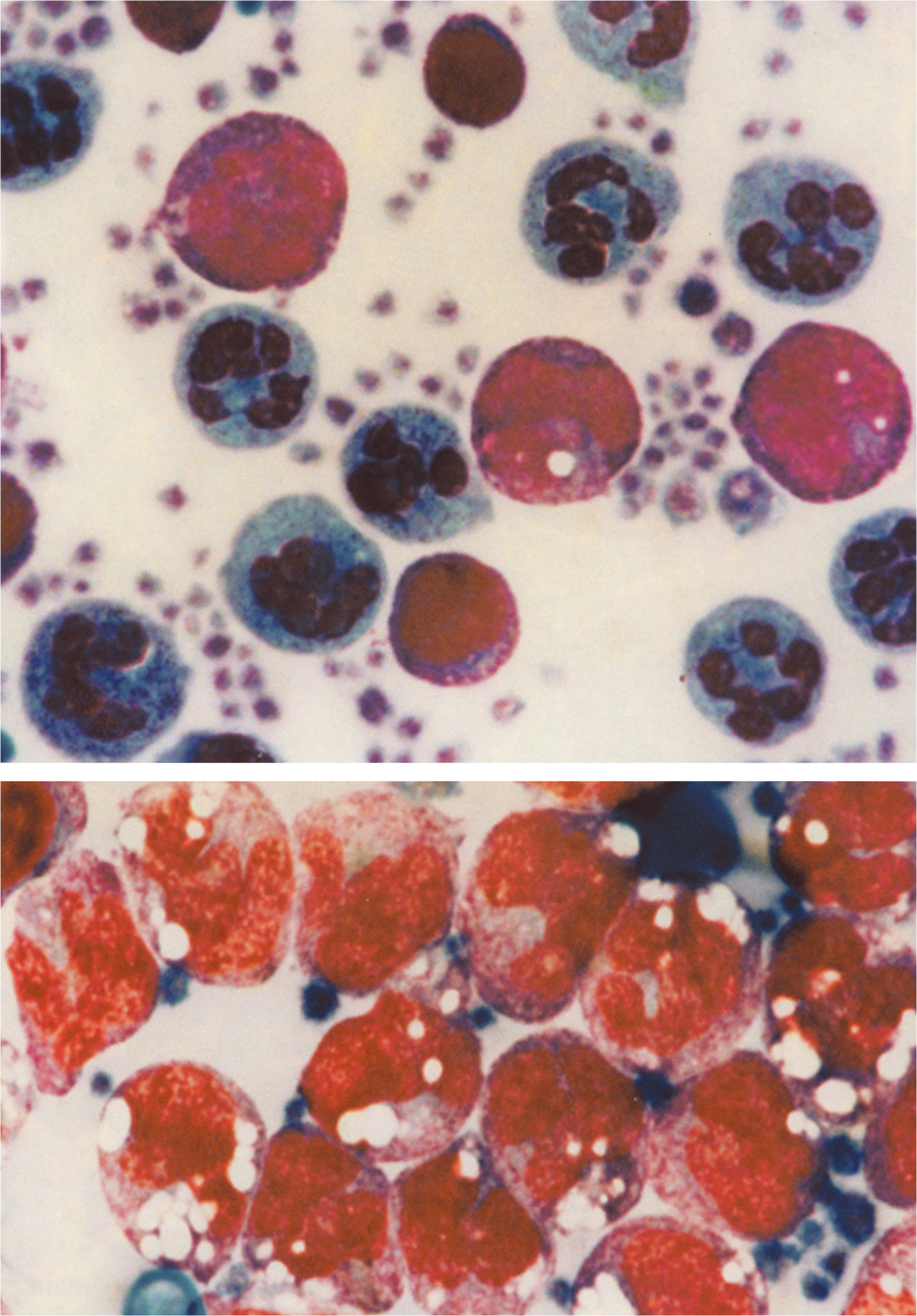
- Chronic lymphocytic leukemia: Using PANOPTIKON or RAPIDIFF, details of nuclei and cytoplasm are distinct. With LYMPHOCOLOR, lymphocytes usually stain yellow, implying their B cell origin in most cases.
- Monocytic leukemia: MONOCOLOR imparts a violet color to the cytoplasm of leukemic monocytes. LYSOCOLOR selectively stains lysosomes in some leukemic monocytes and myeloblasts in this disorder.
- Immune thrombocytopenic pupura: With PANOPTIKON or RAPIDIFF, nuclei and cytoplasm of megakaryocytes are brightly colored and precisely delineated. Using MEGACOLOR, the cytoplasm of these megakaryocytes stains a distinctive magenta color.