A versatile stain for all hematopoietic cells.
Intended Use :
Using PANOPTIKON as a single agent stain, differential coloration of blood and bone marrow cells can be produced. Except for eosinophil granules which stain bright turquoise blue and erythrocytes with stain orange green, the colors produced by PANOPTIKON are virtually identical to those obtained with conventional panoptic stains, like Wright's or Giemsa's. However, PANOPTIKON produces brighter colors than the conventional stains and gives superior visualization of nuclear and cytoplasmic structures such as nuclear chromatin, nucleoli and granules. Since PANOPTIKON is a single agent, it does not require the complex mixtures of eosin, methylene blue, azures, and unstable eosinates found in traditional stains.
Principle :
PANOPTIKON utilizes a highly metachromatic substantially pure dye, capable of imparting a plurality of colors to blood and bone marrow cells, thereby facilitating their identification.
Reagents :
1. PANOPTIKON stain in absolute methanol.
2. Buffer.
Procedure :
1. Flood slides or coverslips containing peripheral blood, bone marrow, or buffy coat with PANOPTIKON for 5 minutes. The presence of undissolved dye particles in the stain solution does not affect the performance of the stain.
2. Without washing, add an equal amount of buffer to the PANOPTIKON stain covering the slide or coverslip and stain for 5 minutes. Drain off liquid from slide or coverslip by touching the edge to a piece of filter paper.
3. Wash by grasping the slide or coverslip with a forceps, and vigorously agitating in a beaker containing buffer for 15 seconds.
4. Blot on filter paper and mount with xylene soluble synthetic resin mounting medium.
Results :
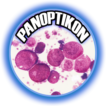
Peripheral blood and bone marrow cells stain colors that are virtually identical to those found with traditional panoptic stains, except that with PANOPTIKON, the colors are more intense and the delineation of cellular structures unusually precise and clear. Particularly, this is seen in nuclear chromatin, nucleoli, and cytoplasmic granules. In contrast to traditional stains like Wright's, PANOPTIKON produces bright turquoise blue granules in eosinophils. As with conventional stains, polychromasia appears as a pale lavender color. Malaria parasites, trypanosomes, erythrocytic inclusions such as basophilic stippling and Howell-Jolly bodies can also be distinguished with PANOPTIKON, and stain similarly to that found with Wright's or Giemsa's stain.
This versatile stain is particularly valuable for demonstrating cytoplasmic and nuclear features of granulocytic cells, as well as cells of erythroblastic and megakaryocytic derivation. Cytoplasmic granulation is stained intensely in both normal and abnormal granulocytic cells, and nuclear features are distinct.
Panoptikon Kit Includes:
1 - 4 oz. Stain
1 - 4 oz. Buffer
$60.00 per kit
~ sufficient for 60 slides or 120 coverslips


