A stain for T helper and T cytotoxic/ suppressor lymphocytes.
Intended Use :
IMMUNOCOLOR is intended to differentially stain T helper lymphocytes and T cytotoxic/suppressor lymphocytes. Based upon differences in color, it is possible to distinguish between the two types of lymphocytes using the ordinary light microscope. As a result, IMMUNOCOLOR serves as a rapid microscopic screening test for T helper and T cytotoxic/suppressor cells, and may help to identify those samples requiring further study by other methods including flow cytometry.
Principle :
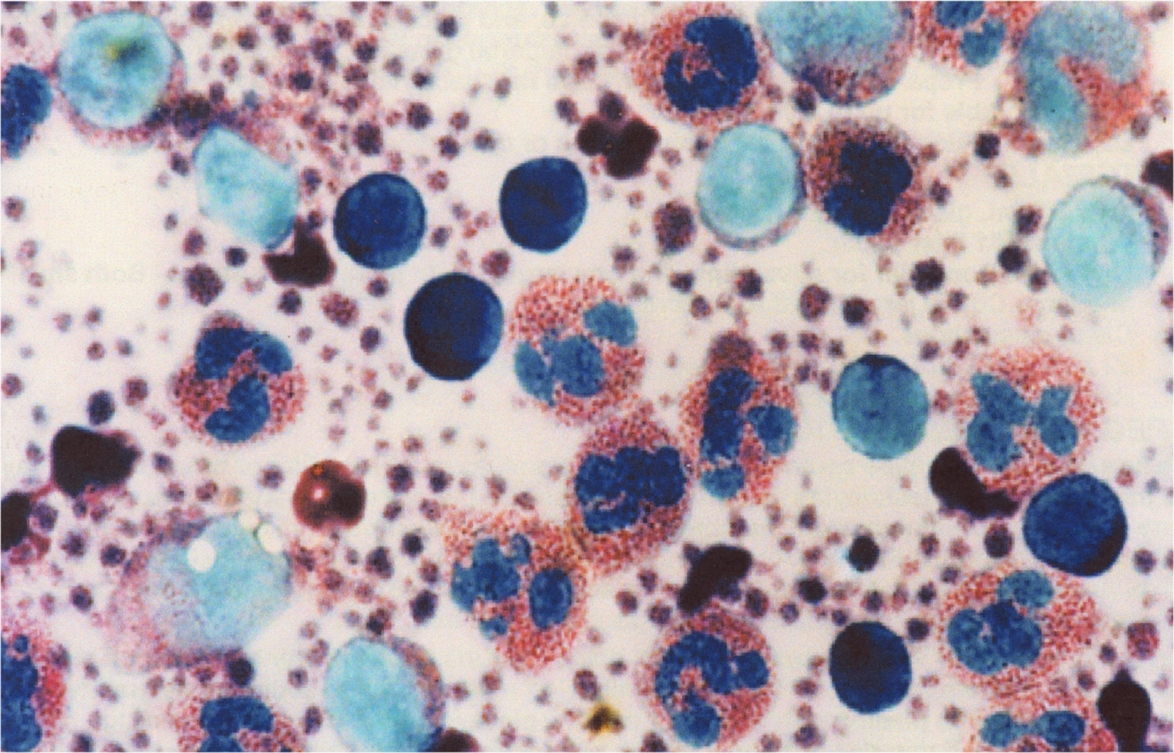
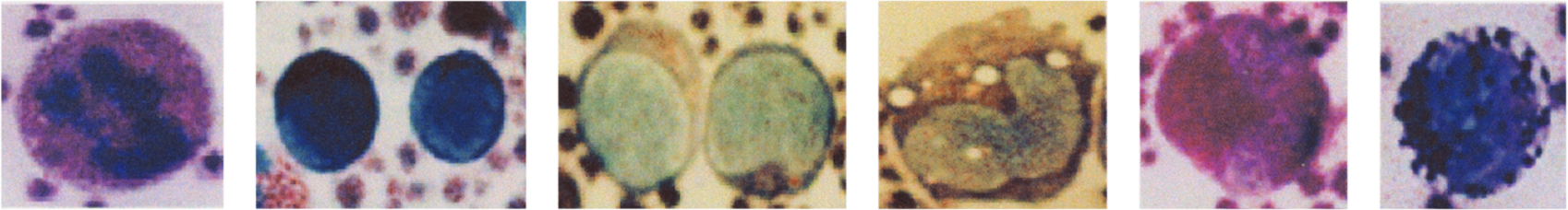
Exposure of lymphocytes first to a methanolic solution of a metachromatic basic cationic dye and then to an aqueous solution of the same dye leads to differential coloration of lymphocytes. Based on a unique dye chemistry, IMMUNOCOLOR stains the nuclei and cytoplasm of T helper cells (CD4) bright royal blue. Simultaneously, IMMUNOCOLOR stains the nuclei and cytoplasm of T cytotoxic/ suppressor (CD8) cells pale green, and the cytoplasm often contains a cluster of maroon colored granules.
Reagents :
1. Solution A: a methanolic solution of a basic cationic dye.
2. Solution B. an aqueous solution of the same basic cationic dye used in Solution A.
3. Tris maleate buffer concentrate 200 mM/L, with a pH 7.6 value.
4. IMMUNOCOLOR rinse buffer, prepared by emptying the contents of the tube labeled IMMUNOCOLOR RINSE BUFFER CONCENTRATE into a 1 L screw top glass or plastic bottle. The 100X buffer concentrate appears turbid, and will become clear after reconstitution. Add 10 mL distilled or deionized water to the empty tube, agitate the tube, pour contents into the bottle, and add 980 mL distilled or deionized water to the bottle. Add 10 drops liquid phenol to the buffer, and store in the refrigerator at 5 degrees C. Prepared in this way, the buffer is stable for one year.
Materials :
1. Two 100 mL glass or plastic beakers.
2. Whatman #1 filter paper.
3. One small Coplin jar for cover slips and one large Coplin jar for glass slides. Both small and large Coplin jars should have screw tops.
4. Cleaned glass slides.
5. 22 x 22 mm and 22 x 40 mm cleaned glass cover slips.
6. Resin based xylene soluble mounting medium.
Specimen Preparation :
Make smears of peripheral blood buffy coat, bone marrow, or lymph node imprints on cleaned glass cover slips or slides and air dry. To make buffy coat preparation, obtain 7 mL peripheral venous blood in an evacuated tube containing EDTA and allow erythrocytes to sediment by gravity for 2 hours or until sedimentation appears complete. Remove leukocyte rich plasma with a Pasteur pipette, place in a separate plastic test tube, and centrifuge for 5 minutes at 2500 rpm (750 x g) at room temperature. Remove all but 100 microliters of the supernatant plasma with a separate Pasteur pipette, resuspend, and make buffy coat smears or cover slips. Alternatively, buffy coats can be obtained by centrifugation of whole blood and removal of the buffy coat layer pipette.
Staining Procedure:
1. Place 50 mL of the buffer solution into a 100 mL glass beaker. This solution can be kept covered at room temperature, and used repeatedly for 10 slides or 20 cover slips. When these numbers are exceeded, the buffer should be discarded and fresh buffer used.
2. Prepare working Solution B by placing 2 mL solution B in a plastic test tube or small flask and to it add 2 mL of tris maleate buffer concentrate. Addition of the buffer to Solution B causes the solution to change from royal blue to dark blue purple.
3. Place slides or cover slips on a staining pan, and flood their surface with 5 mL Solution A. Each time that a fresh rinse buffer solution is used, designate one cover slip or slide as an "initiator," since for maximal color development, the rinse buffer requires a small amount of stain in it before subsequent slides or cover slips are washed. Discard the first slide.
4. After 5 minutes, flood the same slides with approximately 2.0 mL of working Solution B. Addition of this solution to Solution A already covering the slides causes an admixture of the two solutions, and activation of the stain.. A golden sheen may occur transiently on the surface of the liquids, upon addition of working Solution B to Solution A.
5. After 10 minutes, grasp the slide or cover slip with a forceps, tip to the side allowing excess stain to run off, and rinse by vigorous agitation in the beaker containing IMMUNOCOLOR rinse buffer for only 5 seconds.
6. Immediately turn face down on a piece of filter paper, blot dry, then turn face up and air dry. Mount with the cyanacrylate adhesive contained in the kit, by squeezing a drop of adhesive onto the surface of a clean glass slide, and placing the stained coverslip face down. In the case of stained glass slide, mount with a cleaned coverslip in the same way. Binding of the coverslip to the glass slide is virtually immediate and permanent. Take care that the adhesive does not touch the skin, mucous membranes, or eyes.
Results :
For peripheral blood samples, the best results are obtained with buffy coats or leukocyte rich suspensions. Under the light microscope and enhanced by removal of any blue filter and substitution of a didymium filter, T helper cells (CD4) display dark blue nuclei and cytoplasm. In contrast, T cytotoxic/suppressor cells (CD8) show pale green nuclei and cytoplasm, and often 10 to 20 bright maroon colored granules clustered in one area. Neutrophils contain scarlet granules, basophils display bright blue granules and eosinophils granules are large and appear deep maroon. Erythrocytes are blue gray and platelets demonstrate many maroon and purple granules.
Because of differences in the color of the T helper and T cytotoxic/suppressor lymphocytes, it is possible to perform a lymphocyte differential cell count using the IMMUNOCOLOR stain. With IMMUNOCOLOR and a manual cell counter, one can estimate the number of T helper cells as a fraction of the total number of lymphocytes counted (for example, 100 or 200 lymphocytes in a buffy coat preparation), or to estimate a T helper:T cytotoxic/suppressor ratio in the specimen. Estimations of this kind may help to identify which samples require further study by other methods such as flow cytometry.
FOR RESEARCH USE ONLY AND NOT FOR USE IN HUMAN DIAGNOSTICS
Immunocolor Kit Includes:
1 - 2 oz. Solution A
2 - 2 oz. Solution B
1 - 2 oz. Tris Maleate Buffer
1 Super Glue
$60.00 per kit
~ sufficient for 60 slides or 120 coverslips