A stain for monocytes.
Intended Use :
With this two step stain, monocytes in specimens of blood or bone marrow display a distinctive deep purple cytoplasm and red nucleus. Other types of blood and bone marrow cells do not show this type of coloration. As a result, identification of monocytes is improved. This staining reaction avoids the complex unstable incubation mixtures and sensitized couplers found in conventional enzymatic stains for monocytes.
Principle :
Exposure of unfixed blood or bone marrow cells first to a methanolic solution of a basic azo dye, followed by admixture with an aqueous alkaline solution of an oxazine dye, and concluded with a brief rinse in an acidic phosphate buffer, yields an intense purple reaction product in the cytoplasm of normal and leukemic monocytes.
Reagents :
1. Solution A, a methanolic solution of a basic azo dye.
2. Solution B, an aqueous solution of an oxazine dye.
3. Tris Maleate buffer concentrate pH 7.6 200 mM/L.
4. Working solution B, prepared just before use by adding 2.0 mL tris maleate buffer to 2.0 mL solution B. Agitate gently, and the color changes from royal blue to blue purple. This solution is stable at room temperature for 24 hours.
5. Phosphate buffer crystals. To prepare the working buffer at pH 5.6, place all of the crystals in a 1 L flask and add 10 drops of phenol as a preservative. The buffer is stable at 5 degrees C in the refrigerator for 12 months. Do not use the buffer if the solution shows obvious contamination.
Procedures :
1. Make cover slip or smear preparations of peripheral blood, buffy coat, or bone marrow and air dry. Cytocentrifuge preparations and imprints can also be used.
2. Place cover slip or slide in a staining rack, and flood surface with 1.5 mL Solution A.
3. After 5 minutes, add 2.0 mL of working solution B directly on top of the solution A, covering the surface of the slide or cover slip. The act of addition effects the admixture of the two solutions. The presence of undissolved dye particles does not affect the performance of the stain.
4. After 10 minutes, grasp the cover slip or slide with a forceps, tip to the side to allow excess stain to run off, touch the edge to a piece of filter paper, and wash by vigorous agitation in a beaker containing the pH 5.6 phosphate buffer for exactly 5 seconds.
5. Drain dry rather than blot dry, and mount with a synthetic resin based mounting medium on cleaned glass slides for light microscopy. To enhance the purple color of monocyte cytoplasm, substitute a didymium filter for the blue filter covering the light source of the microscope.
Results :
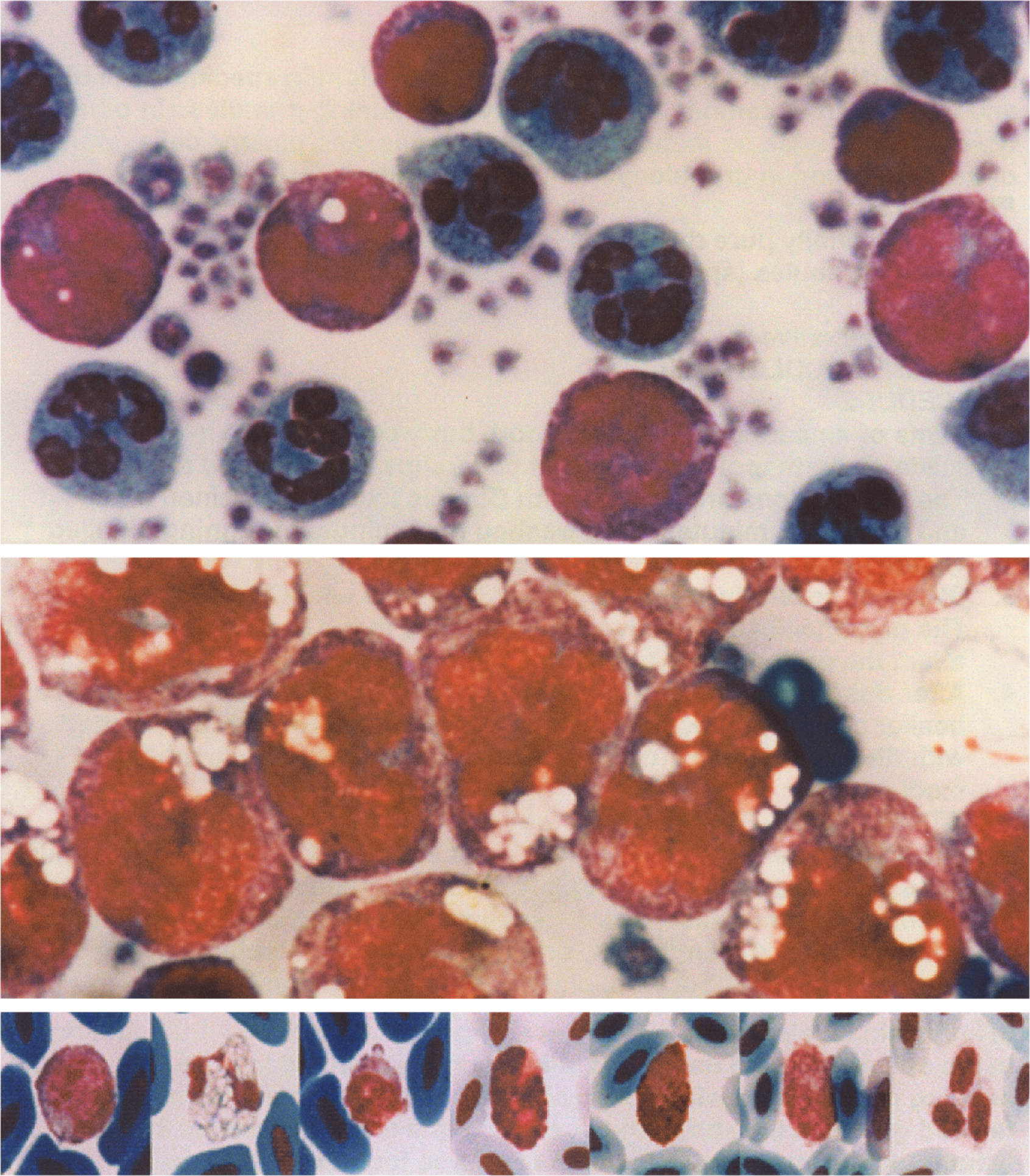
Neutrophils show blue green cytoplasm with a few red granules. Eosinophils contain green granules, and basophils have orange granules. Lymphocytes display deep blue cytoplasm. In all these cells, nuclei stain red brown. Erythrocytes stain blue, and platelets stain pale violet.
In normal monocytes, the cytoplasm stains intensely purple. Often there are pink and magenta granules and purple fibrillar structures scattered throughout the cytoplasm. The nucleus stains a distinctive bright red color that differs from the color of nuclei in other leukocytes in a sample of blood.
In patients with acute myeloblastic leukemia (M1 or M2 in the FAB classification), leukemic blasts show deep blue cytoplasm and pale pink nuclei. Leukemic monocytes from acute monocytic leukemia as typified by the M4 or M5 variants display deep purple cytoplasm and red nuclei.
Monocolor Kit Includes:
1 - 2 oz. Solution A
2 - 2 oz. Solution B
1 - 2 oz. Tris Maleate Buffer
1 - Tube Phosphate Buffer
$60.00 per kit
~ sufficient for 60 slides or 120 coverslips