A stain for mature and immature granulocytes.
Intended Use :
Using GRANULOCOLOR, cells of granulocytic origin in specimens of blood and bone marrow demonstrate intensely red staining granules. In other types of cells, such as monocytes, lymphocytes, and plasma cells, granules of the type found in granulocytic cells are not identified.
Principle :
This substantially pure dye stains both primary granules (lysosomes) and secondary granules (specific granules) in all maturational stages of granulocytic cells.
Reagents :
1. Bouin's fixative.
2. GRANULOCOLOR stain.
Materials :
1. Films or smears of peripheral blood, buffy coat, or bone marrow are made on methanol cleaned glass coverslips or slides and air dried.
2. The preparations are flooded with Bouin's fixative contained in the kit and fixed for 5 minutes, then washed for 1 minute in running distilled water.
3. Slides or coverslips are stained for 3 minutes with GRANULOCOLOR. The Presence of undissolved dye particles in the stain solution does not affect the performance of the stain.
4. After this period, preparations are washed with vigorous agitation in a beaker containing distilled water for 30 seconds to 1 minutes, blotted with filter paper, and mounted with resin based synthetic mounting medium.
5. 22 x 22 mm and 22 x 40 mm cleaned glass cover slips.
Results :
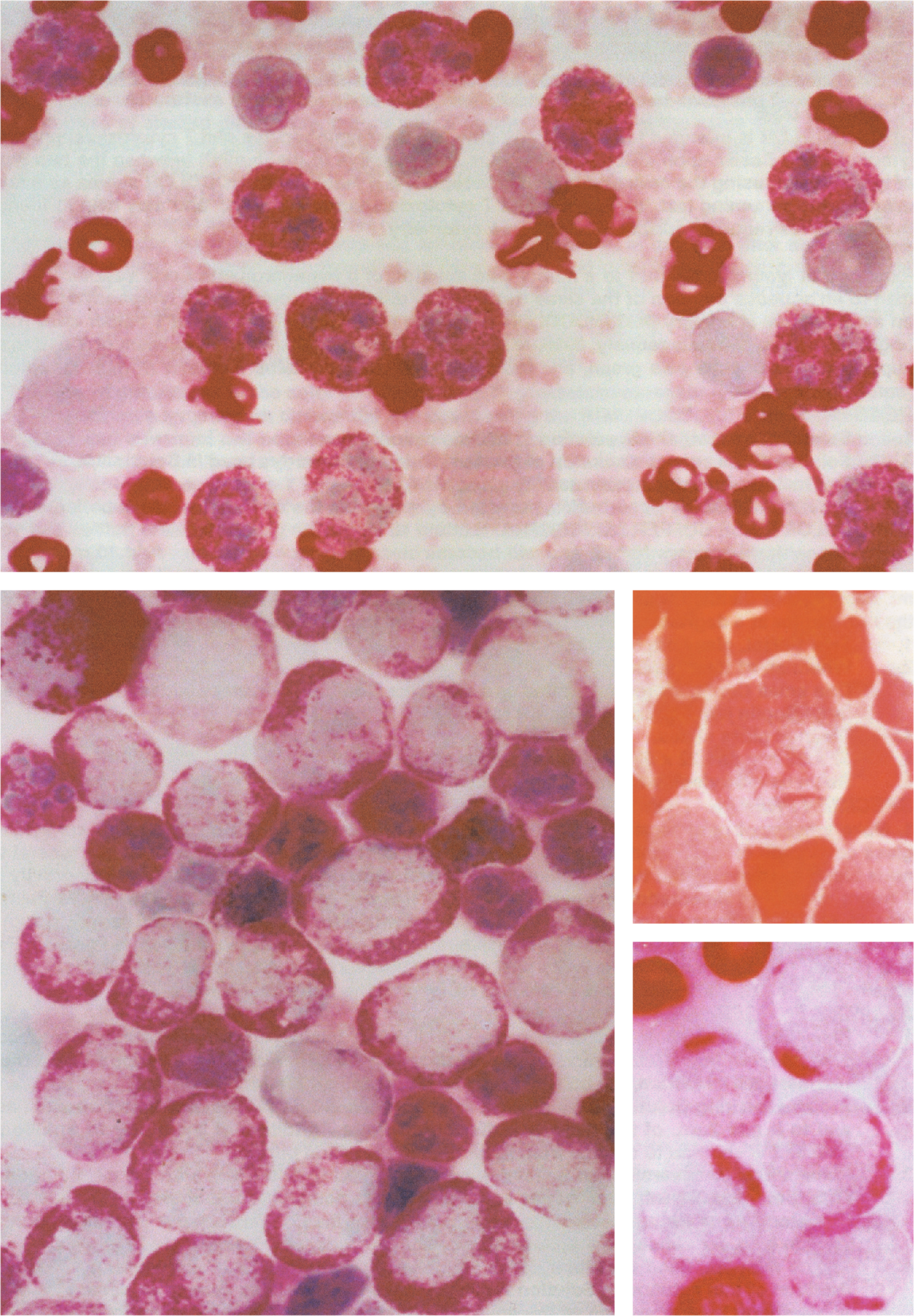
In neutrophils, granules are numerous and stain bright red. In eosinophils, granules stain deep red. In basophils, granules stain red. In lymphocytes and monocytes, granules are not visualized. In bone marrow, granules are identified in cells of granulocytic origin, and are not detectable in megakaryocytes, lymphocytes, plasma cells, or fibroblasts.
In leukemic myeloblasts, 2-20 red staining granules can often be observed in the cytoplasm, and usually parallel the reaction for myeloperoxidase and the staining pattern seen with Sudan black B. In myelomonocytic leukemia, few red granules may be found in some of the leukemic blasts. In acute lymphoblastic leukemia, granules of the type found in leukemic myeloblasts are not identified. GRANULOCOLOR can be used as a substitute for the specific esterase reaction (naphthol AS-D chloroacetate esterase), since it stains granules in both mature and immature granulocytic cells. GRANULOCOLOR stains Auer rods in leukemic myeloblasts and promyelocytes bright red.
Granulocolor Kit Includes:
1 - 4 oz. Stain
1 - 4 oz. Bouins Fixative
$60.00 per kit
~ sufficient for 120 slides or 240 coverslips