
A stain for T helper (CD4) cells.
Intended Use :
The CeeDee4 stain is intended for the rapid and selective staining and visualization of CD4 (T helper) lymphocytes in specimens containing blood or bone marrow cells.
Principle :
CeeDee4 selectively stains CD4 (T helper) lymphocytes an intense deep violet color. This color is distinctive, and other types of lymphocytes such as CD8 (cytotoxic/suppressor) cells, as well as other types of blood and bone marrow cells, do not display this type of coloration. In most instances, these other cells are either pale or unstained. Used in the manner described, the CeeDee4 stain permits a rapid visualization of CD4 lymphocytes and thereby a screening for and estimation of their numbers in the stained specimen using an ordinary light microscope.
Reagents :
1. CeeDee4 stain.
2. FAA fixative.
3. Rinse buffer, prepared by emptying the tube containing buffer crystals into a 1 L plastic or glass graduated cylinder. Wash any remaining crystals adherent to the interior of the tube into the graduated cylinder with distilled or deionized water, bring volume to 1 L with distilled or deionized water, agitate to dissolve, and place in a screw top glass or plastic 1 L bottle. Add 1 mL liquid phenol to retard bacterial growth and place in the refrigerator at 5 degrees C. Prepared this way and provided that bacterial growth does not occur, the buffer is stable for up to 1 year.
4. Distilled or deionized water, for rinsing slides or cover slips after fixation in FAA.
Materials :
1. Two 100 mL glass or plastic beakers.
2. Whatman #1 filter paper.
3. One small Coplin jar for cover slips, and one large Coplin jar for glass slides. Both should have screw tops.
4. Clean glass slides.
5. 22 x 22 mm and 22 x 40 mm cleaned glass cover slips.
6. Resin based xylene soluble mounting medium.
Specimen Preparation :
Glass slides or cover slips containing peripheral blood, as in a blood film or blood smear, may be used. Buffy coat leukocytes smeared on a glass slide or cover slip may be utilized. To prepare buffy coat, obtain one 7 mL sample of peripheral venous blood by venipuncture in an evacuated tube containing EDTA and centrifuge at 2500 rpm (750 x g) at room temperature for 10 minutes. Using a Pasteur pipette, remove and discard supernatant plasma, and carefully remove buffy coat layer and place into a separate 10 x 75 mm tube.
Alternatively and for improved yield of cells, place the tube containing anticoagulated blood upright at room temperature for 2-3 hours, allowing erythrocytes to sediment by gravity. Remove leukocyte rich plasma with a Pasteur pipette, place in a plastic conical centrifuge tube, and centrifuge for 5 minutes at 2500 rpm (750 x g) at room temperature. Remove all but approximately 100 microliters of the supernatant plasma, and resuspend the button of leukocytes and platelets by agitation on a vortex mixer. Using this concentrated leukocyte suspension, make cover slip films or slide smears and air dry. Coverslip preparations or slides of bone marrow particles and imprints of lymph nodes on cover slips or slides may also be used. These preparations should be air dried prior to fixation in FAA as described below.
Procedure :
1. Using a small Coplin jar for coverslips and a large Coplin jar for glass slides, fix the specimens in FAA fixative contained in the kit for 5 minutes at room temperature. Use Coplin jars that have a screw top. Apply the screw top to retard evaporation.
2. After 5 minutes, remove the slide or coverslip with a forceps, agitate vigorously in a beaker containing distilled water for 15 seconds, and place face down on a piece of filter paper. Blot dry and place face up for further drying in the air.
3. Place 100 mL rinse buffer into a 100 mL beaker. After use, the buffer can be covered with paraffinized film, kept at room temperature, and used repeatedly for 20 glass slides or 40 coverslips before the buffer must be discarded and replace with fresh buffer.
4. Place coverslips or slidesin Coplin jar containing CeeDee4 stain. For buffy coats, stain for only 1 minute. For peripheral smears or peripheral blood coverslip films, stain for 10 minutes. For lymph node imprints and bone marrow aspirates, stain 1 minute.
5. After the staining time has elapsed, grasp the slide or coverslip with a foreceps and remove from the Coplin jar containing the stain. Touch the slide or coverslips to a piece of filter paper to remove excess stain, and rinse by vigorous agitation in the rinse buffer solution for exactly 5 seconds. Place slide or coverslipupright on a piece of filter paper, leaning against the Coplin jar, and drain dry rather than blot dry. For repeated use of the stain, screw on the tops of the Coplin jar to retard evaporation.
6. Using synthetic resin based xylene soluble maounting media, mount stained coverslips onto the surface of cleaned glass slides. If stained glass slides are used, mount with a cleaned 22x40mm coverslip. View under an ordinary light microscope.
Results :
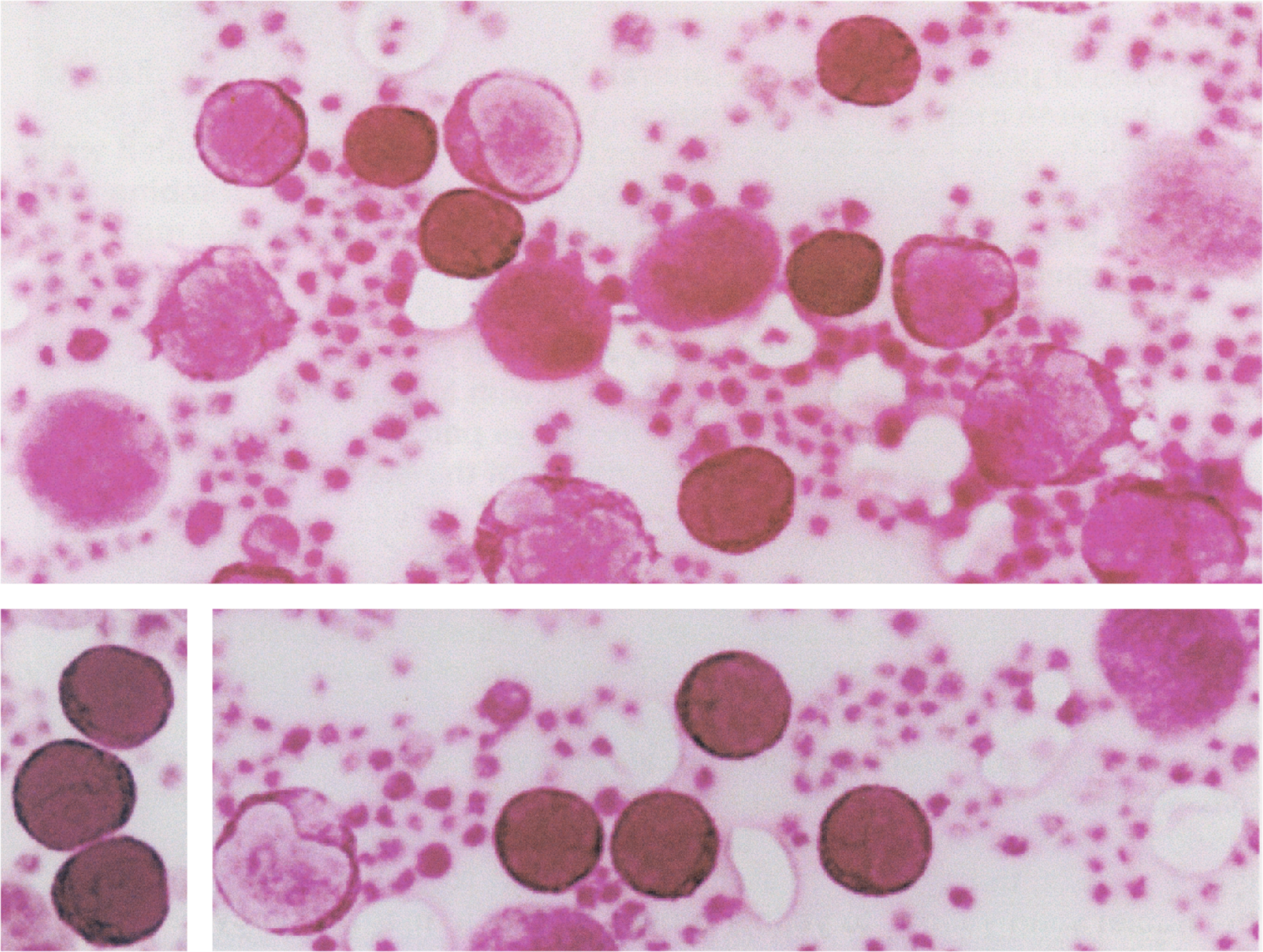
For peripheral blood samples, the best results are obtained with buffy coats or leukocyte rich suspensions. Stained in the manner described, and correlated with purified lymphocyte populations obtained from a cell sorter, CD4 (T helper) lymphocytes display an intense deep violet color of the nuclei and cytoplasm. Often a network of purple colored fibrillar structures are seen in the nuclei. In contrast, CD8 (cytotoxic/suppressor) lymphocytes show pale if any staining of the nucleus, and faint lavender staining of the cytoplasm. Neutrophils show faint lavender staining of nuclei and cytoplasm, monocytes stain faint lavender and may contain one or more clear vacuoles, granules in eosinophils are unstained, and granules in basophils stain faint orange. Erythrocytes and platelets are not stained. in specimens of bone marrow, nuclei of erythrocytes and plasma cells stain pale lavender.
FOR RESEARCH USE ONLY AND NOT FOR USE IN HUMAN DIAGNOSTICS
CeeDee4 Kit Includes:
1 - 2 oz. Stain
1 - 2 oz. FAA Fixative
1 - Tube Phosphate Buffer Salts
$60.00 per kit
~ sufficient for 120 slides or 240 coverslips